Figure 10.

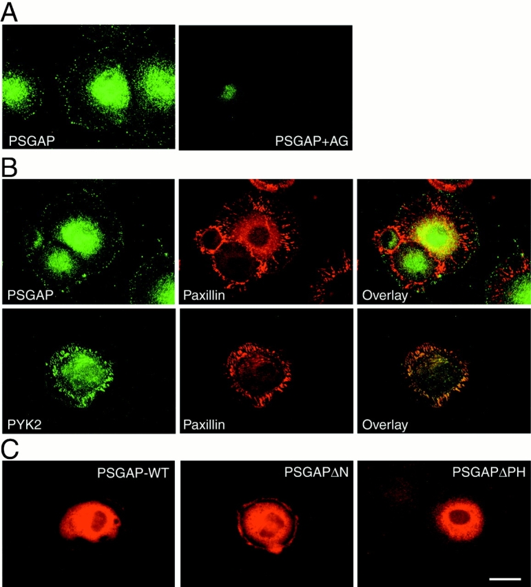
Expression of PSGAP at the cell periphery in 10T1/2 fibroblasts. 10T1/2 cells were fixed with 4% paraformaldehyde for 20 min, blocked with 10% BSA, and immunostained with indicated antibodies. (A) Immunostaining using antibodies against PSGAP or antibodies preabsorbed with the GST-PSGAP antigen. (B) Coimmunostaining with anti–PSGAP or anti–PYK2 and antipaxillin antibodies. PSGAP and PYK2 were visualized by FITC-conjugated secondary antibodies, whereas paxillin appeared in red with rhodamine-conjugated secondary antibodies. (C) Immunostaining of PSGAP wild-type (PSGAP-WT), NH2-terminal–deleted PSGAP (PSGAPΔN), and PH-domain–deleted PSGAP (PSGAPΔPH) in transfected 10T1/2 fibroblasts. Cells were transfected with the indicated constructs and stained with anti–PSGAP antibodies. Bar, 50 μm.
