
Figure 2.
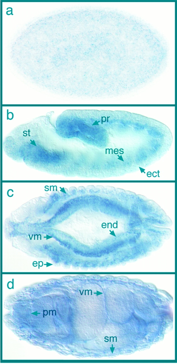
Expression of Drosophila ILK mRNA during embryogenesis. (a) Lateral view of a cellular blastoderm embryo showing low levels of ILK mRNA distribution in the whole embryo. Anterior is left in this and all subsequent panels. (b) Lateral view of an embryo during gastrulation (stage 11), dorsal side up. Staining is seen in the mesodermal layer (mes) underlying ectoderm (ect), including the stomodeum (st) and the proctodeum (pr). (c) Dorsal view of an embryo at stage 13 showing high levels of ILK mRNA in the progenitors of the visceral muscles (vm) and somatic muscles (sm). Lower levels of expression can be detected in endodermal origin midgut (end) and in epidermis (ep). (d) Dorsal view of an embryo at stage 16. Strong expression of ILK mRNA is visible in the visceral muscles (vm), somatic muscles (sm), and pharyngeal muscles (pm).