
Figure 6.
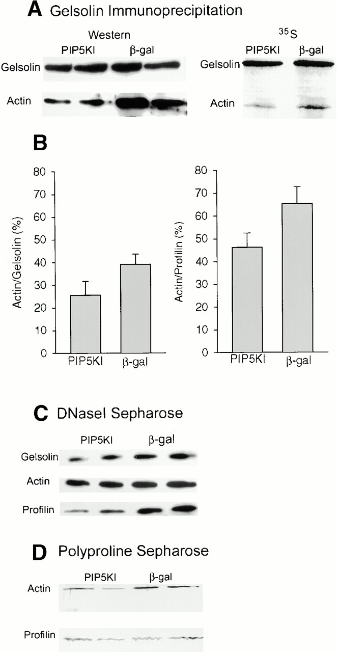
Effects of PIP5KI overexpression on the interaction of gelsolin and profilin with actin. (A) Gelsolin immunoprecipitation. Cells were labeled with 35S-translabel and gelsolin was immunoprecipitated. Gelsolin and actin were detected by Western blotting (left) or by autoradiography (right). (B) Quantitation of actin:gelsolin and actin:profilin ratios after immunoprecipitation. Gelsolin was immunoprecipitated from 35S-labeled cells as in A, and the intensities of the actin band in the autoradiogram was expressed as a percentage of that of gelsolin. Profilin was pulled down with poly-l-proline sepharose. Profilin and actin was detected by Western blotting, and the intensity of the actin band was expressed as a percentage of that of profilin. Values shown are mean ± SEM of four experiments for gelsolin and two for profilin. (C) Actin pull down with DNaseI-sepharose. Actin and associated gelsolin and profilin were detected by Western blotting. (D) Profilin pull down with poly-l-proline sepharose. Proteins were detected by silver staining.