
Figure 5.
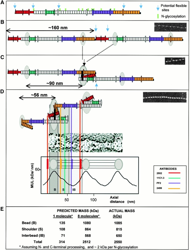
A model of fibrillin alignment in microfibrils. Schematic diagram depicting the folding of fibrillin molecules in a beaded microfibril. (a) A single NH2- and COOH-terminal processed molecule and N-glycosylation sites are indicated. Antibody epitopes are colored on the molecule (red, 2502; blue/cyan, 11C1.3; purple, PF2; orange, 2499). (b) Alignment in 160-nm periodicity microfibrils with dashed circles representing regions of the molecule predicted to contribute to the bead. (c) Molecular folding that would generate ∼100-nm periodicity; solid black lines show the position of possible transglutaminase cross links and solid circles show the bead position. (d) Packing and folding that would generate the stable 56-nm periodicity microfibrils. The evidence for the model is presented in the Discussion. While the predicted folds are shown, it should be noted that the precise packing arrangement of folded segments contributing to the bead remains unresolved.