
Figure 4.
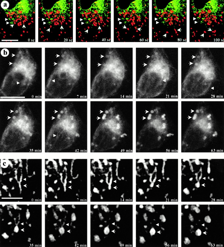
Real-time microscopy. CDV are immobile structures formed by maturation processes in some specific regions of the ER. a, Living GFP-tagged cavDGV-transfected cells were incubated in a medium containing a marker for acidic compartments, lysoTracker. Cells expressing GFP-cavDGV were selected under the microscope and the focus was kept constant. Images of both channels (green/red) with a delay of 10 s between channels were captured over 5 min. The figure shows a selected sequence of 6 consecutive frames (20-s interval between images) representing a total period of 100 s. LysoTracker-labeled vesicles showed a rapid bidirectional motility (arrows) but cavDGV–GFP positive vesicles showed no significant motility in any axis (arrowheads). b and c, In an attempt to capture the formation of CDVs, cells were transfected with GFP-tagged cavDGV for 18 h and were then observed by video microscopy. Cells were selected for the presence of forming rings (see arrows in the first panel) and the focus was kept constant during the entire experiment. Images were captured with a constant interval of 7 min. The figure shows a selected sequence of 10 consecutive frames representing a total period of 63 min. At time 0, cavDGV–GFP was abundant in the tubular elements of the ER and in the Golgi area. Progressively the protein is less abundant in the ER and Golgi complex and accumulates in the forming rings (arrows) to shape well-defined CDV ring-like structures in ∼60 min. c shows a selected high magnification example of CDV formation. The protein can be detected moving in waves along the tubules of the ER (arrowheads) until the formation of the DGV-containing vacuolar structures (arrows). Bars: (a and b) 5 μm; (c) 1 μm. Supplemental videos are available at http://www.jcb.org/cgi/content/full/152/5/1057/DC1.