
Figure 8.
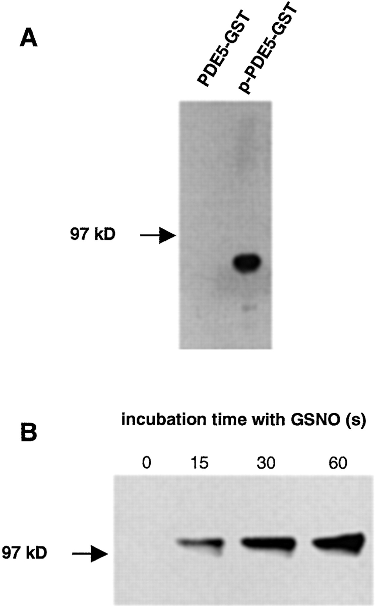
Detection of PDE5 phosphorylation. (A) Purified unphosphorylated and phosphorylated GST–PDE5 fusion proteins (∼85 kD; 200 ng, each) were subjected to SDS-PAGE and transferred to nitrocellulose. Membranes were incubated with the antiphospho-PDE5 antibody in a dilution of 1:10,000. (B) Platelets were stimulated with 300 μM GSNO. At the indicated time points, aliquots (3.6 × 10⁷ platelets) were removed and proteins were separated by SDS-PAGE and transferred to nitrocellulose. Membranes were incubated with phospho-PDE5–specific antibody in a dilution of 1:50,000.