
Figure 3.
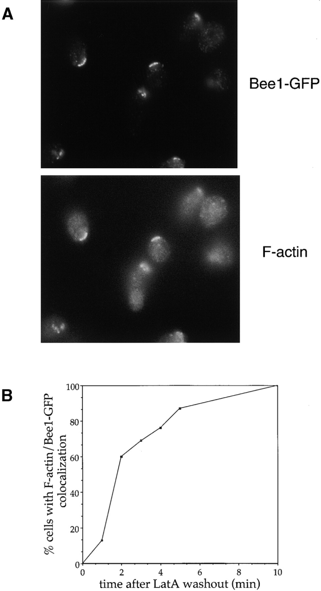
Polarized Bee1-GFP defines the site of actin polymerization in vivo. (A) A population enriched in polarized unbudded cells was generated by releasing G1-arrested cells into the cell cycle in the presence of Lat-A. 1 h after release, Lat-A was washed away, and cells were fixed at various time points. The cells were stained for Bee1-GFP (anti-GFP antibody) and F-actin (rhodamine phalloidin). Shown are cells from the 5 min time point. (B) Quantitation of results from the experiment described in A. At various time points after Lat-A washout, the percentage of cells with F-actin colocalizing with polarized Bee1-GFP was determined. Only cells with polarized Bee1-GFP were counted.