
Figure 8.
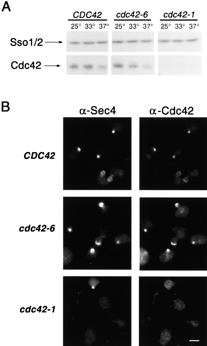
Levels of Cdc42 accumulation and localization of Cdc42 and Sec4 in CDC42 , cdc42-6 and cdc42-1 strains. (A) Whole cell lysates were prepared and subjected to SDS-PAGE analysis and then blotted with anti-Cdc42 or anti-Sso2 antibodies. (B) Immunofluorescence probing for Sec4 and Cdc42 localization was done after a shift to the restrictive temperature of 33°C for CDC42 (top), cdc42-6 (middle) and to 37°C for cdc42-1 (bottom). Bar, 5 μm.