
Figure 4.
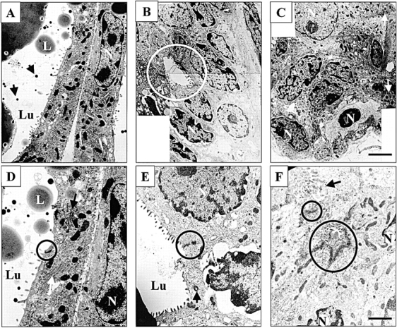
Disorganized structures of Stat5- and PrlR-null epithelia. Stat5- and PrlR-null mammary epithelia at parturition were analyzed by electron microscopy. (A and D) Control mammary epithelium at lactation day 1 was fully differentiated and contained secreted milk proteins and lipid droplets. Golgi apparatus (white long arrow) and RER (white long arrow) were detected. Lu, lumen; L, lipid droplet; N, nucleus; black short arrow, β-casein micelles; black circle, tight junction. (B and E) Alveoli-like structures of PrlR-null epithelium were more organized than Stat5-null epithelia. Lumina were detected in alveolar-like structure (white circle). The centrosome was located close to the apical membrane (black arrow). Tight junctions were maintained (black circle). (C and F) Alveoli-like structures in Stat5-null epithelia were disorganized and cell–cell contacts were aberrant (black arrow). Frequently, two or more pseudo-lumina were detected in one alveolar-like structure (black circles). The cells near the basement membrane contained lipid droplet-like structures (white arrow). Active Golgi apparatus and a RER in Stat5- and PrlR-null epithelia were not apparent. Bars: (A–C) 2.4 μm; (D–F) 1.6 μm.