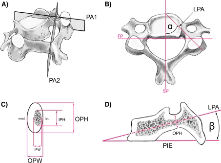
Fig. 1.
Mid-cervical vertebra showing cuts and lines used for the CT measurements: aPA 1 vertical cut through the longitudinal pedicle axis (LPA), PA 2 vertical cut through the isthmus of the pedicle, perpendicular to PA 1. b Superior view: sagittal plane (SP), frontal plane (FP), longitudinal pedicle axis (LPA), pedicle transverse angle α (PTA) between PA 1 and SP. c Cut PA 2 through the pedicle isthmus: outer pedicle height (OPH), outer pedicle width (OPW), inner pedicle height (IPH), inner pedicle width (IPW). Note that the lateral wall is always thinner than the medial one. d Cut PA 1: plane of the inferior vertebral endplate (PIE), pedicle sagittal angle β (PSA) between the plane of the inferior endplate (PIE) and longitudinal pedicle axis (LPA)

