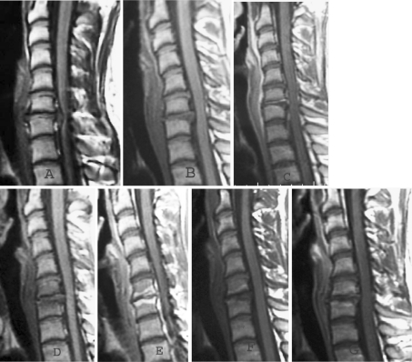
Fig. 4.
Pre-operative T1 weighted MRI sagittal image (a) of a 23-year-old female patient presented with 3 month history of radiculopathy and myelopathy, showing disc prolapse at C6/7 level. Compared to first post-operative day pre contrast T1 weighted image (b), the post contrast T1 weighted image (c) has shown no enhancement of vertebral end-plates at operated level. At 6 weeks, compared to pre contrast image (d), the post contrast image (e) has shown brilliant enhancement of end-plates and adjacent vertebral marrow. But at 6 months, compared to pre contrast image (f), the post contrast image (g) has shown low grade enhancement in vertebra marrow at operated level

