
Fig. 1.
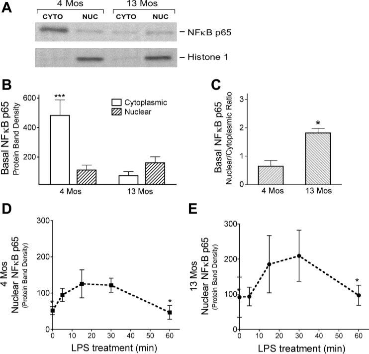
NF-κB p65 protein in cerebral blood vessels from 4- and 13-mo-old ovariectomized (OVX) rats. A and B: nuclear (nuc) and cytoplasmic (cyto) fractions were obtained from vessels isolated from whole brains of 4- and 13-mo-old animals to determine basal levels of NF-κB p65 protein. A: representative Western blot for NF-κB p65; histone 1-immunopositive bands verify the nuclear fractionation. B: NF-κB p65 band densities, normalized to α-actin (means ± SE; n = 4 rats). ***Significantly different from 3 other groups. C: ratio of NF-κB p65 protein levels in nuclear and cytoplasmic fractions (n = 4). *Significantly different from 4-mo-old animals. D and E: nuclear translocation of NF-κB following ex vivo LPS treatment of pial vessels freshly dissected from the brains of 4- (D) and 13-mo-old (E) OVX rats. Densities of NF-κB p65 protein bands (normalized to α-actin) from Western blots of vascular nuclear fractions are shown for different times of LPS treatment. Values are means ± SE; n = 4. *Significantly different from 20- and 30-min time points, P ≤ 0.05.