
Fig. 3.
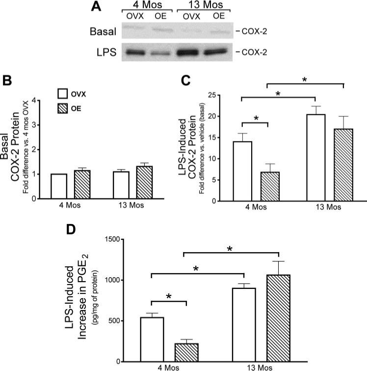
Effects of age and estrogen on LPS induction of cyclooxygenase-2 (COX-2) protein and PGE2 production in cerebral blood vessels. A–C: COX-2 protein in cerebral blood vessels isolated from whole brains of 4- and 13-mo-old OVX and OE rats 6 h after in vivo administration of vehicle (A and B, basal levels) or LPS (A and C). A: representative Western blot. Mean band densities are expressed as the fold increase in protein relative to the level in vessels from either 4-mo OVX (B) or vehicle-treated (C) rats analyzed on the same Western blot. D: PGE2 production from pial vessels freshly dissected from 4- and 13-mo-old OVX and OE animals. Vessels were incubated in vitro with LPS or vehicle for 6 h. The LPS-stimulated increase in PGE2 production was calculated as the difference in PGE2 levels in the vessel incubation medium in the absence or presence of LPS. Means ± SE are plotted; n = 5. *Significantly different from each other in the same group, P ≤ 0.05.