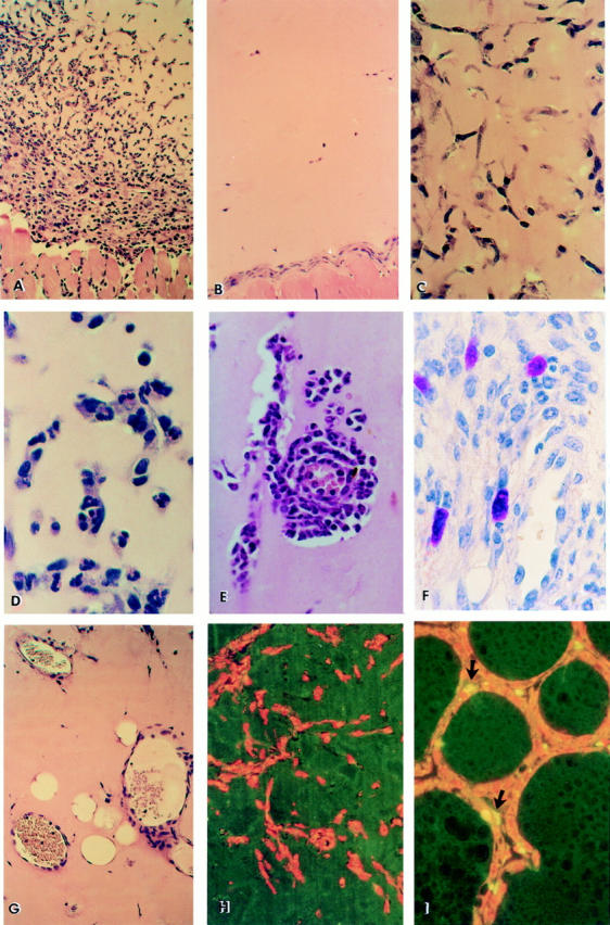
Figure 1.

Histological and immunohistochemical analysis of Matrigel implants. Representative hematoxylin-eosin–stained section of Matrigel containing 5 μg/ml anti-Fas mAb Jo2 (A) implanted in wild-type mice and excised 6 d after injection (6 mice per condition in each experiment). Progressive invasion of cells into the Matrigel implant and formation of capillaries is observed (×200). (B) Control: mAb Jo2 was replaced by hamster IgG (×200). Similar results were obtained by using nonagonistic anti-Fas mAb RMF6 or by injecting Matrigel with mAb Jo2 into lpr Fas-mutant mice (not shown). Kinetic of anti-Fas mAb Jo2-induced inflammatory angiogenesis (×400); (C) Initial migration of endothelial cells aligning to form cord-like structures at day 1; (D) appearance of PMN inside the lumen of neocapillaries (day 2); (E) marked extravasation of PMN throughout the neoformed capillary into the Matrigel (day 6); (F) metachromatic staining by Toluidine blue of mast cells infiltrating Matrigel at day 6 (×600); (G) formation of canalized vessels containing erythrocytes and leukocytes (day 4–6). Immunohistochemical detection of apoptotic cells by the TUNEL method (normal nuclei are counterstained with propidium iodide in red); (H) Section of Matrigel with anti-Fas mAb Jo2 at day 6, displaying total absence of apoptotic cells among the infiltrating cells (×200). The same results were obtained on Matrigel excised 6 h, 12 h, 24 h, 5 d, and 10 d after injection (not shown); (I) Rat regressing mammary glands obtained at the fourth day after weaning showing apoptotic cells (arrows) along the acini, as positive control (×200).
