
Figure 4.
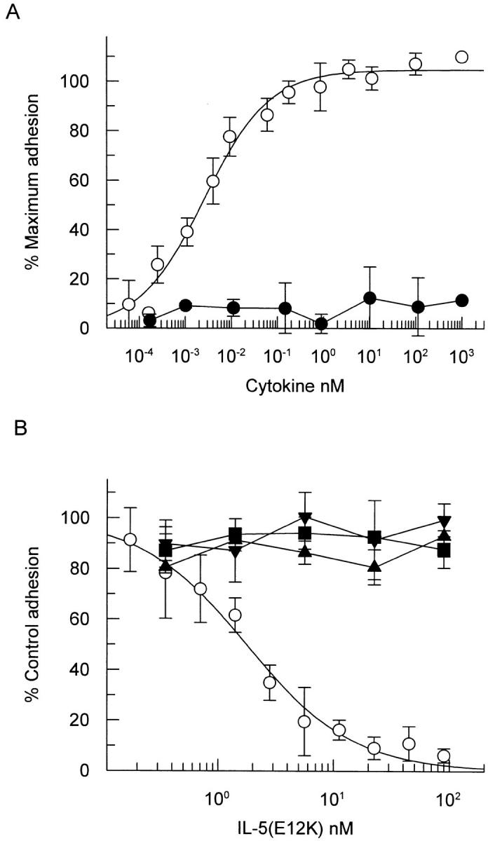
IL-5 (E12K) exhibits no agonist activity (A) and is a specific IL-5 antagonist (B) in a cytokine induced eosinophil activation assay. (A) Human eosinophils were incubated with increasing concentrations of either wild-type (open circles) or IL-5 (E12K) (closed circles) for 30 min at 37°C, in a 96-well microtiter plate precoated with human IgG. After a washing step, the adherent eosinophils were lysed and the endogenous peroxidase activity measured in a colorimetric assay. (B) In antagonist experiments eosinophils were incubated with either 20 pM IL-5 (open circles), 180 pM IL-3 (closed squares), 6 pM GM-CSF (closed triangles), or 1,000 pM TNF-α (inverted triangles) in the presence of increasing concentrations of IL-5 (E12K). The concentrations of wild-type cytokines represent ∼ED80's in the eosinophil adhesion assay. In both panels each value represents the mean ± SEM of three independent experiments using different blood donors. 100% values for IL-5–, IL-3–, GM-CSF–, and TNF-α–induced adhesion were 0.56 ± 0.07, 0.76 ± 0.05, 0.41 ± 0.07, and 0.49 ± 0.06, respectively. Unstimulated levels were 0.15 ± 0.05.