Figure 2.

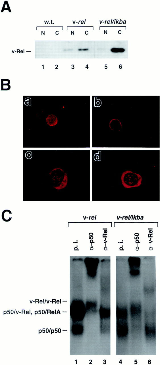
Change of nucleus/cytoplasm distribution of v-Rel in v-rel/ ikba double transgenic thymocytes. (A) Western blot analysis. Cytoplasmic (C) and nuclear (N) fractions from thymocytes and Western blot assays were performed as previously described (31). (B) Immunofluoresence. Thymocytes from 6-wk-old v-rel (a) and v-rel/ikba (b) transgenic mice were spun onto slides, incubated with anti–v-Rel antibodies, and visualized with donkey anti–rabbit Texas red–labeled antibodies. T cells from spleen-bearing tumors of v-rel/ikba double transgenic mice were spun onto slides, incubated with anti–v-Rel (c) or anti-IκBα (d) antibodies, and visualized with donkey anti–rabbit Texas red–labeled antibodies. (C) Decreased p50/v-Rel κB-binding activity in v-rel/ikba double transgenic thymocytes. Electrophoretic mobility shift assays were performed as previously described (23) using nuclear protein extracts from v-rel and v-rel/ikba double transgenic thymocytes. Lanes 1 and 4 were treated with preimmune sera (p.i.), lanes 2 and 5 were treated with anti-p50 antibody (α-p50), and lanes 3 and 6 were treated with anti–v-Rel antibody (α-v-Rel).
