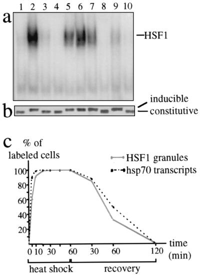
Figure 5.
HSF1 granules and the activation of the heat shock response. Electrophoretic mobility-shift assay (a) and Western blot analysis (b) were performed on whole-cell extracts prepared from HeLa cells after various treatments: 37°C (lane 1), 1 hour at 42°C (lane 2), 1 hour at 42°C followed by 2 hours recovery at 37°C (lane 3), 1 hour at 42°C followed by 6 hours recovery at 37°C (lane 4), 1 hour at 42°C followed by 2 hours recovery at 4°C (lane 5), 1 hour at 42°C followed by 6 hours recovery at 4°C (lane 6), azetidine (5 mM) for 4 hours at 37°C (lane 7), azetidine (5 mM) for 4 hours at 4°C (lane 8), cadmium (30 μM) for 2 hours at 37°C (lane 9), and cadmium (30 μM) for 2 hours at 4°C (lane 10). (c) HSF1 granules were detected by immunofluorescence in HeLa cells, and hsp70 gene transcription was followed by detection of the nascent hsp70 transcripts by FISH. The percentages of cells displaying HSF1 granules (gray line) and hsp70 transcription sites (dashed line) were measured at different time points during exposure at 42°C and recovery.

