
Figure 4.
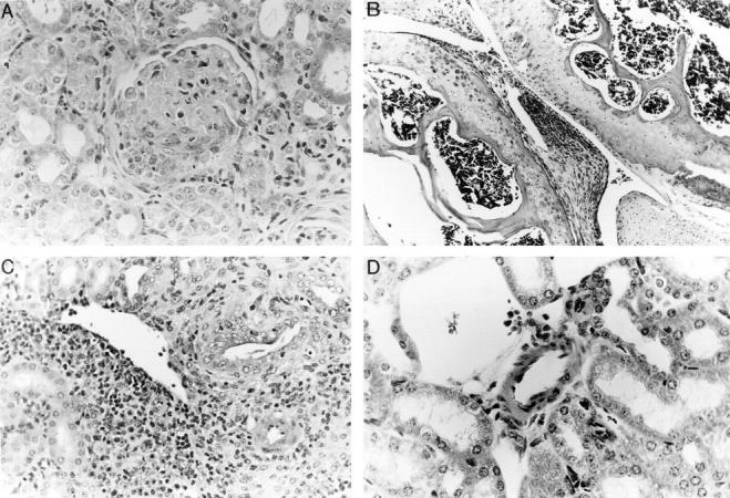
(A, B, C, and D) Histology of tissues from NOS2-modified MRL–lpr/lpr mice. 20-wk-old mice were examined. A shows a representative section from kidney of a (−/−) MRL–lpr/lpr mouse, demonstrating glomerulonephritis with inflammation, sclerosis, and crescent formation. Comparable lesions were observed in (+/+) mice and in (+/−) mice. B shows a representative section of a knee joint from a (−/−) MRL–lpr/lpr mouse. There is synovial proliferation and inflammation. Comparable lesions were observed in (+/+) mice and in (+/−) mice. C and D show kidney sections containing medium-sized arterioles from MRL–lpr/lpr (+/+) (C) and (−/−) (D) mice. That from the (+/+) a mouse in C demonstrates vasculitis, whereas that from a (−/−) mouse in D is essentially normal with no evidence of vasculitis. (Hematoxylin and eosin stain; original magnifications 200×).