
Figure 3.
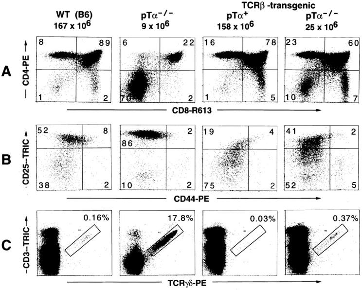
The effects of TCR-β transgenes on various aspects of thymic T cell development in the absence of pTα (the right most panel in A, B, and C). A TCR-β-transgenic mouse expressing functional pTα (TCR-β-transgenic, pTα+), a wild-type C57BL6 (WT [B6]), and a nontransgenic pTα-deficient (pTα−/−) mouse are included as controls. (A) CD4/CD8 profiles. The figures on top of each panel give the total number of thymocytes found in the particular mouse analyzed in this experiment. These values are very typical for mice with the respective genotype as seen in many similar experiments. (B) Analysis of triple-negative (CD3−CD4−CD8−) thymocytes for differential expression of the developmental markers CD25 and CD44. Thymocytes from three mice of the same genotype were pooled. CD4- and CD8-expressing cells (single positive and DP thymocytes) from B6 and TCR-β-transgenic, pTα+ mice were complement depleted (see Materials and Methods) before staining. For cytofluorometric analysis, thymocytes were incubated with an antibody mix containing CD4–FITC, CD8–FITC, CD3–FITC, GR1–FITC, MAC1–FITC, CD19–FITC (the latter three antibodies are specific for granulocytes, macrophages, and B cells, respectively), CD44–PE and CD25–biotin, and in a second step with streptavidin–Tricolor. FITC-positive cells were excluded from the analysis by electronic gating (data not shown). The cells shown in the four panels are thus highly enriched for immature triple-negative thymocytes. (C) The generation of γδ-expressing cells is suppressed in TCR-β-transgenic mice, even in the absence of pTα. Thymocytes were stained with a PE-conjugated TCR-γδ–specific antibody and CD3–biotin, in a second step with streptavidin–Tricolor. Only thymocytes that are positive for both CD3 and TCR-γδ were considered as genuine γδ-expressing cells (as defined by the rectangular gate). The data shown in A and C were obtained in the same experiment with the same mice.