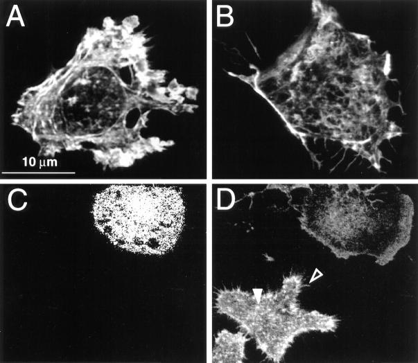
Figure 2.
The effect of microinjected C3 exotoxin on the actin cytoskeleton of FcγRIIA-transfected COS and J774 cells. (A) FcγRIIA-transfected COS cells were mock-injected with the fluorescent dye Lucifer yellow dissolved in injection buffer alone, fixed, and stained with rhodamine phalloidin to visualize F-actin. Note the presence of stress fibers and actin accumulation at adhesion plaques. (B) FcγRIIA-transfected COS cells were injected with C3 exotoxin 60 min before fixation and staining with rhodamine phalloidin. Note the disappearance of stress fibers and focal adhesions and the persistence of F-actin along membrane ruffles. (C) Lucifer yellow emission of J774 cells, identifying the top cell as having been injected with C3 exotoxin. (D) Rhodamine-phalloidin staining of the cells shown in C. Note the decrease in F-actin content, and the absence of filopodia (open arrowhead) and focal complexes (solid arrowhead) in the C3-injected cell (top right) compared to the uninjected (bottom left) cells. Representative of eight separate experiments.

