
Figure 3.
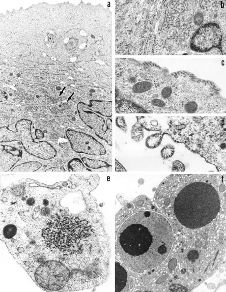
Electron microscopy analysis of MV replication in DC. Transmission electron microscopic analysis performed on day 2 after infection with LYS-1, shows the complete replication cycle of MV in DC. (a) Syncytia containing ⩾10 DC nuclei and cytoplasmic structures typical of the viral nucleocapsid (arrow) (×7,600); (b) A higher magnification of the viral nucleocapsid close to a nucleus (×28,000); (c) Viral glycoproteins identified at sites of membrane rufflings (×34,000); (d) Virus budding leading to the release of intact virions (×46,000); (e) Fragments of DC syncytia resulting from cell clasmatosis, and containing viral nucleocapsids in the cytoplasm (×28,000); and (f) Apoptosis of DC syncytia with condensed chromatin in the nuclei (×8,600).