
Figure 2.
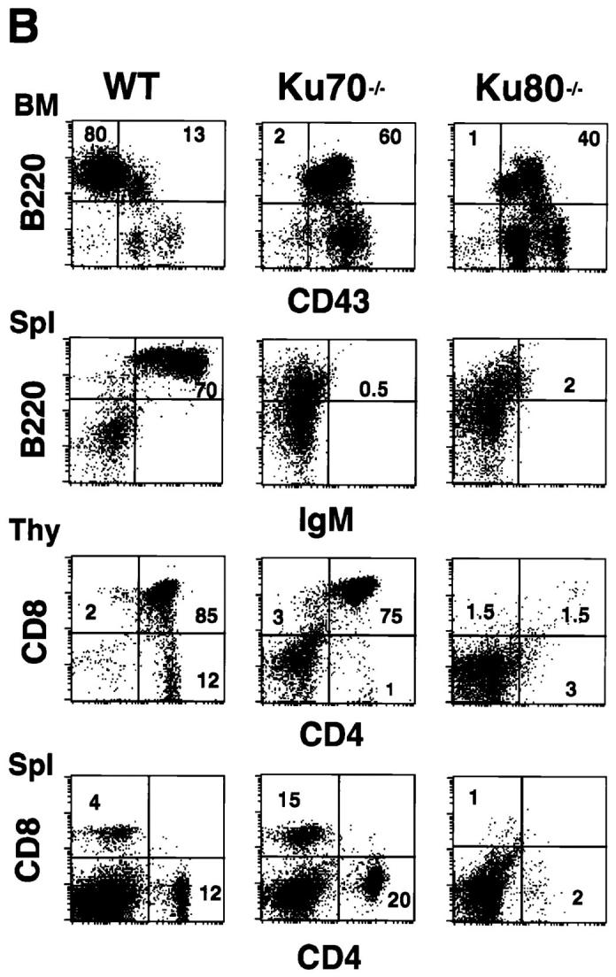

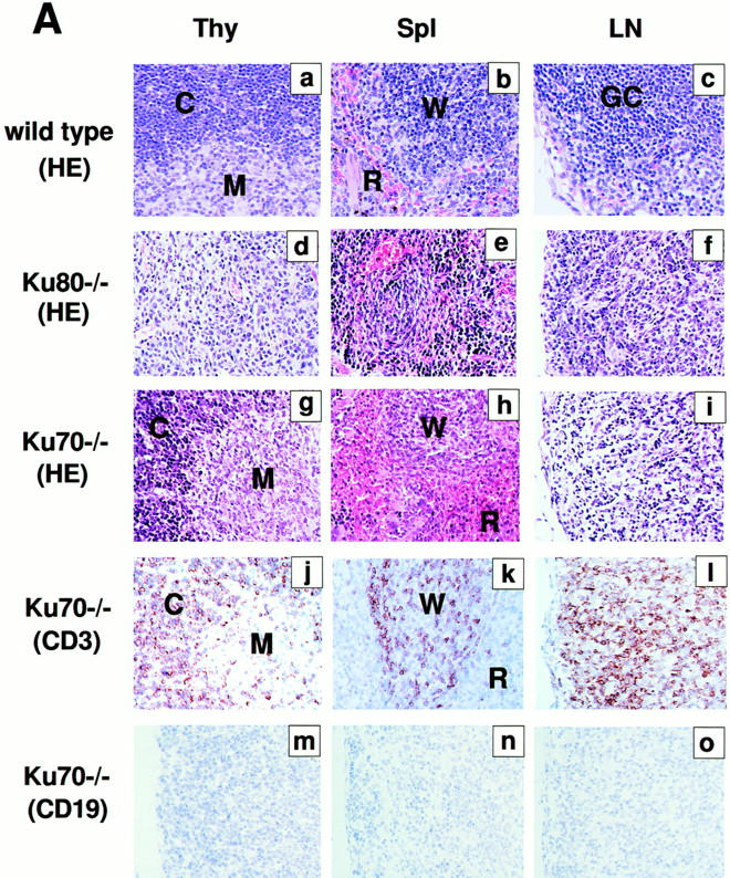
Development of B lymphocytes, but not T lymphocytes, is blocked at an early stage in Ku70− /− mice. (A) Histology of thymus (Thy), lymph nodes (LN) and spleens (Spl) from wild-type control mice, Ku70− /− mice, and Ku80− /− mice (23). Cortex (C) and medulla (M) are indicated. W, white pulp; R, red pulp; GC, germinal center. (a–i) Tissue sections were stained with haematoxylin and eosin (HE); (j– l) tissue sections were stained with anti-CD3 (CD3); and (m–o) tissues were stained with anti-CD19 (CD19). Anti-CD3 and anti-CD19 antibodies were tested in both frozen and paraffin sections of wild-type lymphoid organs and showed the expected specific patterns of staining (data not shown). (B) Flow cytometric analysis of thymocytes (Thy) bone marrow (BM) and spleen (Spl) cells from Ku70− /− mice, Ku70+ /+ littermates, and Ku80− /− mice. CD4, anti-CD4 monoclonal antibody; CD8, anti-CD8 monoclonal antibody; B220, anti-B220 monoclonal antibody; CD43, anti-CD43 monoclonal antibody; IgM, anti-Igμ heavy chain monoclonal antibody. The data were gated for live lymphoid cells based on forward and side scatter properties; 10,000–20,000 cells were analyzed per sample. (C) Analysis of TCR-β chain expression in Ku70− /− mice. Thymocytes and spleen cells were obtained from Ku70− /−, Ku80− /−, and wild-type littermates and analyzed for expression of CD4, CD8, and TCR-β by three-color flow cytometry. The TCR-β expression of both CD4+ and CD8+ SP T cells were shown.