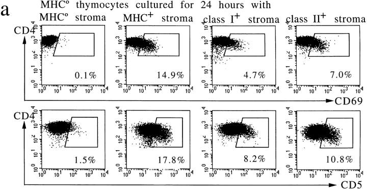
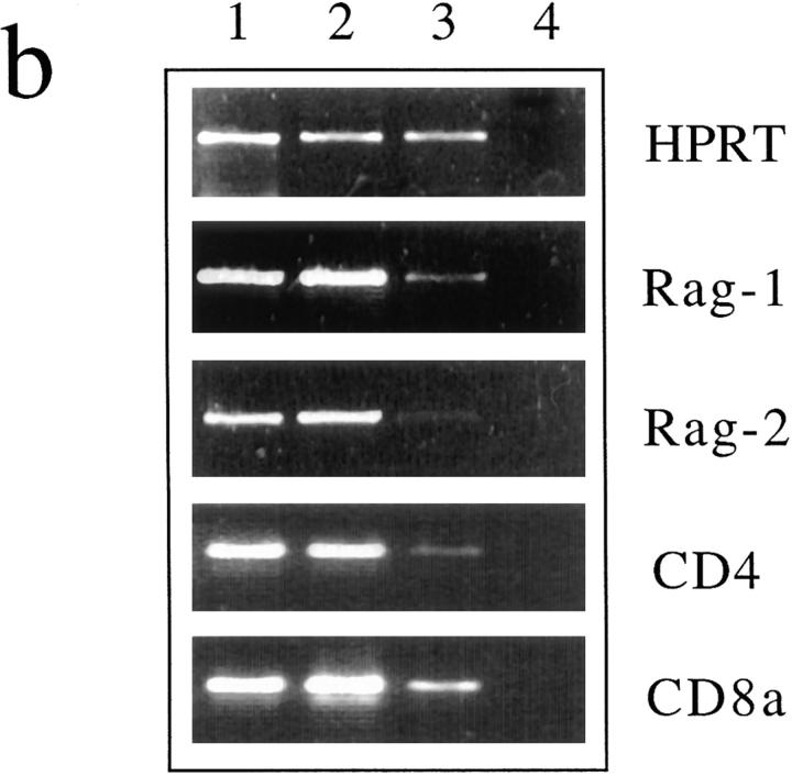
Figure 3.
Preselection thymocyte responses to MHC class I and II are largely additive and, in addition to CD69, involve CD5 upregulation, downregulation of RAG-1 and -2 mRNA, and perturbed CD4 and CD8 expression. (a) Elevated CD5 and CD69 expression by MHC-naive thymocytes 24 h after contact with MHC. Note that responses to class I and II (Ab) are approximately additive. These responses are independent of superantigens, as verified using MHC-expressing cells from mice without mouse mammary tumor virus integrants (provided by Dr. E. Simpson, data not shown). (b) Downregulation of RAG-1 and -2 mRNA 24 h after MHC contact. RT-PCR analysis of thymocytes isolated by fluorescence activated cell sorting from reaggregates with MHC-deficient (lane 1) or MHC+ stroma (lanes 2 and 3). The latter were separated into CD69-negative (lane 2) and -positive cells (lane 3). RAG-1 and -2 signals are barely detectable in CD69-positive thymocytes; CD4 and, to a lesser extent, CD8a mRNA appear downregulated. RT-PCR for HPRT serves as a control; lane 4 contains no cDNA.


