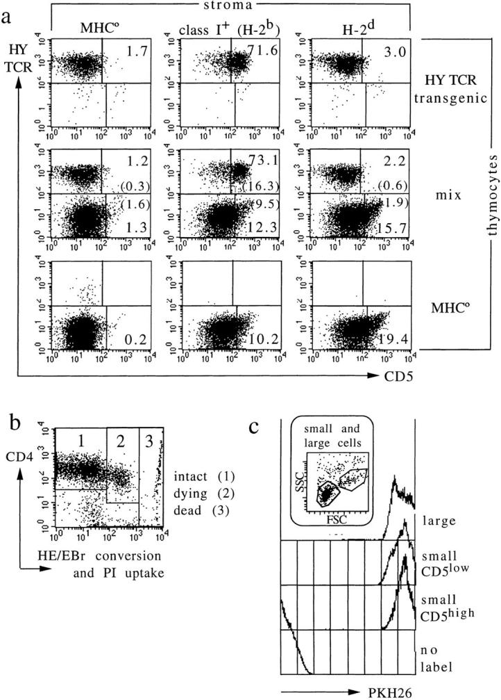
Figure 4.

Bystander activation, unscheduled display of activation markers during apoptosis, or preferential expansion do not account for the frequency of MHC responsive thymocytes. (a) CD5 induction by cognate TCR–MHC interactions. HY TCR transgenic thymocytes from RAG-1–deficient mice lacking MHC class I expression respond strongly to selecting (H-2b), but not to nonselecting (H-2d) MHC molecules (top; Note that in contrast to an earlier study [45], our experiment excludes endogenous TCR rearrangements by using RAG-1–deficient thymocytes). Reaggregates containing mixtures of HY TCR transgenic and MHC-naive, TCR nontransgenic thymocytes (middle) show that both populations respond independently, and without significant bystander activation (compare top and bottom to middle). The percentage of cells in each region is indicated. In the middle panel, the percentage of activated cells among TCR transgenic and nontransgenic thymocytes is calculated separately (percentage of total cells is given in brackets). (b) Most cells recovered from short-term aggregate cultures are viable (i.e., exclude propidium iodide; PI) and metabolically intact, as indicated by low oxidative conversion of hydroethidine (HE) to ethidium bromide (EBr ; reference 33). Displays in Fig. 3 a and data in Table 1 are gated on the region marked 1. (c) MHC-responsive thymocytes do not selectively expand in short-term culture. Thymocytes were labeled with the fluorescent membrane dye PKH26 (Sigma Chemical Co.) before coculture with stroma for 24 h. Control traces are of unlabeled and large (cycling) thymocytes; the latter illustrating dye dilution on cell division. PKH26 fluorescence is the same for MHC-responsive and -nonresponsive cells (defined by CD5 expression level).
