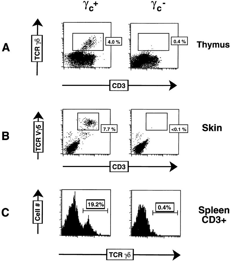
Figure 1.
Flow cytometric analysis of γ/δ T cells from adult γc + and γc − mice. (A) Thymocytes were stained with FITC–anti–pan-TCR-γ/δ (GL3), PE–anti-CD3, biotin-anti–CD4, and biotin–anti-CD8α. CD4− CD8− (DN) cells were electronically gated. (B) Skin-resident DETCs were isolated as indicated in Materials and Methods, and stained with biotin–anti-TCR Vγ5 and FITC–anti-CD3. (C) Splenocytes were monocyte depleted by adherence and stained with FITC–anti–pan-TCR-γ/δ, PE–anti-CD3, biotin–anti-CD4, and biotin–anti-CD8α. CD3+ DN cells were electronically gated. Biotinylated antibodies were revealed with streptavidin-Tricolor.

