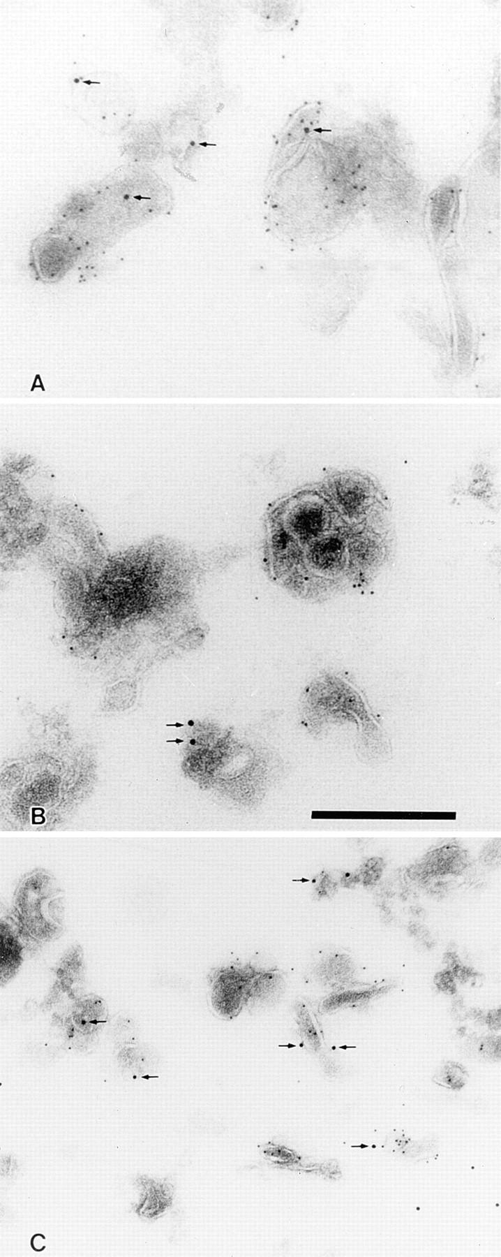
Figure 2.

ImmunoEM localization of BCR-internalized antigen in isolated CIIV as well as endosomes and lysosomes. Bar: 300 nm. (A) ImmunoEM localization of BCR-internalized antigen in isolated CIIV. A20μWT cells were pulsed with antigen (i.e., 20 μg/ml PC–OVA) for 30 min at 37°C and then fractionated by FFE. Isolated CIIV were then analyzed by multiple label immunoEM with rabbit anti-class II and 5 nm protein A–gold followed by rabbit anti-OVA and 10 nm protein A–gold. Isolated CIIV were found to be doubly positive for both class II and BCR-internalized antigen (PC–OVA; arrows), indicating that a portion of the BCR-internalized antigen is delivered to CIIV. The specificity of the staining for class II molecules and BCR-internalized antigen is demonstrated by the lack of label over areas of the section that do not contain vesicular structures. (B and C) ImmunoEM localization of BCR-internalized antigen in isolated endosomes and lysosomes. Endosome/lysosome– containing FFE fractions from PC–OVA pulsed A20μWT cells were double labeled for class II and BCR-internalized antigen as in A. BCR-internalized antigen (PC–OVA; arrows) could be detected in both class II– negative (B) and class II–positive (C) vesicles.
