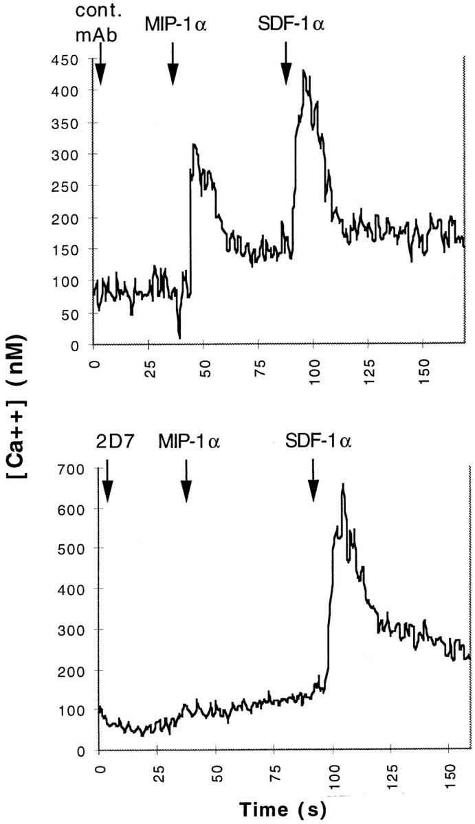
Figure 4.

mAb 2D7 inhibits [Ca2+]i flux in CCR5 L1.2 cells in response to MIP-1α. CCR5 L1.2 cells were labeled with Fura-2 as described in Materials and Methods, and stimulated sequentially with mAb, followed 40 s later with MIP-1α, and 100 s with SDF-1. [Ca2+]i fluorescence changes were recorded using a spectrofluorometer. The tracings were representative of three separate experiments. In the top panel, an irrelevant mAb (MOPC-21) was used, and in the bottom panel, mAb 2D7. Antibodies were used at a final concentration of 20 μg/ml. MIP-1α was used at 100 nM and SDF-1 was used at 200 nM.
