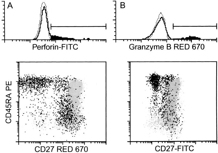
Figure 6.
Expression of mediators of cytotoxicity in CD8+ T cell subsets. After surface staining of freshly isolated CD8+ cells with CD45RA and CD27 mAbs, cells were fixated and permeabilized and intracellularly present perforin and granzyme B were detected with specific mAbs. The histograms show the staining for the intracellular proteins. Dotted lines indicate the negative controls. The positive cell fraction is painted black. The dot-plots show the distribution of positive cells (black) among the total cell population (gray). Note that the different dot-plot staining is due to the use of either biotinylated CD27 mAb in combination with red 670 or FITC-labeled CD27 mAb.

