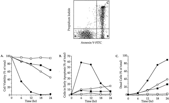
Figure 1.
Time course of apoptosis in isolated thymocytes cultured alone or in the presence of immobilized anti-CD3 mAbs or dexamethasone. The upper panel shows the flow cytometry measurement of annexin V binding (x axis) vs. propidium iodide uptake (y axis) on thymocytes cultured for 12 h in the presence of immobilized anti-CD3. Viable cells and cells in early apoptosis are annexin V− PI− (A) and annexin V+ PI− (B), respectively. Dead cells are annexin V+ PI+ (C). The lower panels display the evolution of cell viability (A), the percentage of cells in early apoptosis (B) and the mortality (C) in thymocyte populations incubated at 4°C (open square) or cultured at 37°C alone (closed squares), in the presence of immobilized anti-CD3 mAb (open circles) or with dexamethasone (closed circles). This experiment produced identical results in a total of four independent occasions.

