Figure 2.
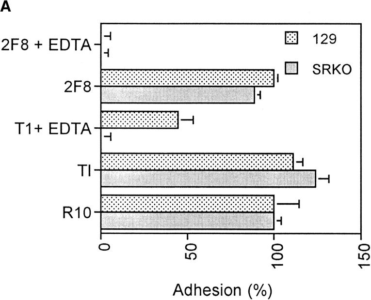
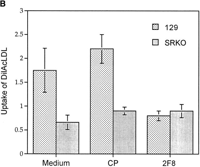
Functional phenotype of BCG-activated SRKO and wild-type Mφ. (A) Adhesion of activated Mφ to FBS-coated TCP. Cells were plated at 3 × 10⁵ macrophages/well of a 96-well plate in the presence of various mAbs and/or EDTA. BCG-recruited peritoneal cells from SRKO and wild-type (129) mice show similar adhesion in medium (R10) alone, but in the presence of both EDTA and isotype-matched control mAb (T1), adhesion of SRKO Mφ is completely inhibited (compare with wild-type 129), showing that SR-A accounts for all EDTA-resistant adhesion. Addition of 2F8 mAb and EDTA is required to completely inhibit the remaining adhesion of 129 activated Mφ. Adhesion is represented as the percentage of that obtained in medium alone, where 100% represents an absorbance (OD) at 450 nm of 0.15. Results (mean ± SD) are derived from a single experiment (n = 4) and are representative of three similar experiments. (B) The uptake of DiIAcLDL by SRKO and wild-type (129) BCG-recruited peritoneal cells. Cells were harvested as in A and the uptake of DiIAcLDL measured as described in Materials and Methods. SRKO endocytose only 40% of the DiIAcLDL taken up by 129 cells. 2F8 mAb reduces the uptake by 129 cells to that of SRKO cells, whereas an isotype-matched control Ab (CP) has no effect. Uptake of DiIAcLDL is expressed by units of fluorescence (mean ± SD) of four replicate wells and is relative to a blank well of cells without the addition of DiIAcLDL. Results are derived from a single experiment and are representative of at least three similar experiments. Uptake of DiIAcLDL by 129 Mφ in the presence of medium or CP is significantly greater than uptake by SRKO Mφ (P <0.005).


