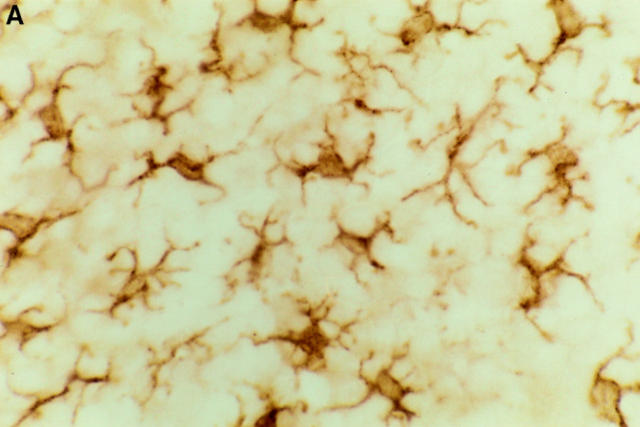
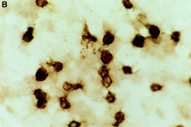
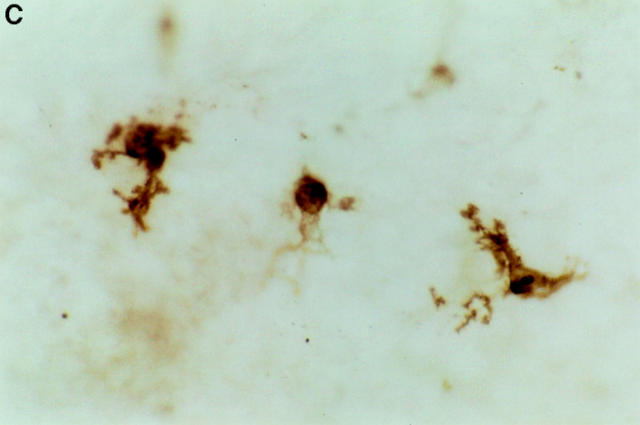
Figure 3.
Immunohistochemical staining of epidermal LCs in situ. Epidermal sheets were prepared either from (A) naive mice, or from skin explants that had been incubated for 72 h on (B) culture medium containing anti-α6 integrin antibody GoH3 at 50 μg/ml, or (C) culture medium alone. LC are stained for MHC class II using indirect immunoperoxidase staining. Note rounded morphology of LCs in anti-α6 integrin antibody– treated explants in comparison with interdigitating morphology of LCs in naive skin. Magnification, ×600.


