Figure 6.
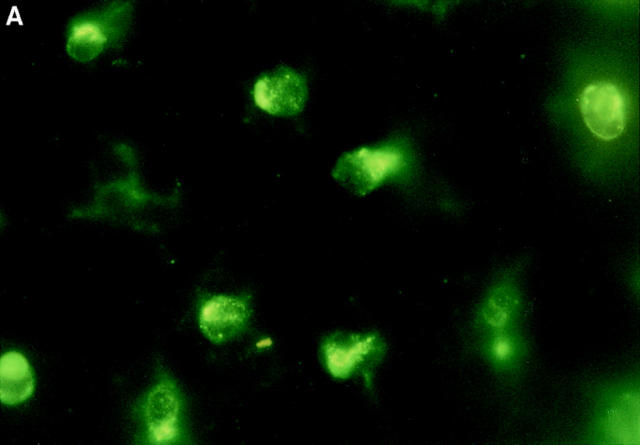
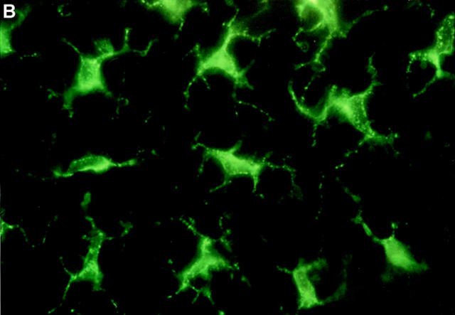
Immunofluorescence staining of epidermal LCs in situ. Groups of mice (n = 2) received a single 100 μl injection intraperitoneally of 40 μg anti-α6 integrin antibody (GoH3) 2 h before intradermal injection into both ear pinnae of (A) 50 ng murine recombinant TNF-α or (B) 0.1% BSA alone. Ears were removed 30 min later, epidermal sheets were prepared, and the morphology of LCs was assessed after indirect immunofluorescence staining for MHC class II expression. Note the rounded morphology of LCs in TNF-α–treated ears in comparison with interdigitating morphology of LCs in controls. Magnification, ×800.


