
Figure 7.
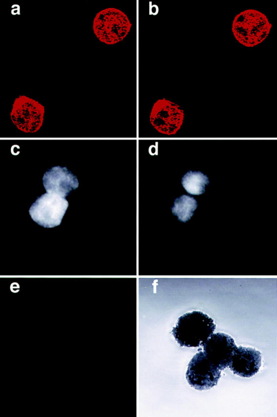
Identification of endogenous ZAP-70 in the nucleus of Jurkat cells by IF staining. Jurkat (a, b, c, and d) and P116 (e and f ) cells were stained with affinity-purified anti–ZAP-70 antiserum. 0.5 μM slices through the middle of the cells are displayed (a, b, e, and f ). Two 0.5 μM slices, 1 μM apart, are shown for anti–ZAP-70–stained Jurkat cells (a and b), and one for P116 cells (e). An image of the same field as in e, using the reflector optics, indicates the specificity of the Ab stain (f ). The nuclear staining of the anti–ZAP-70 is further verified by colocalization of anti– ZAP-70 (c) with the Hoechst DNA stain (d) in the same cells.