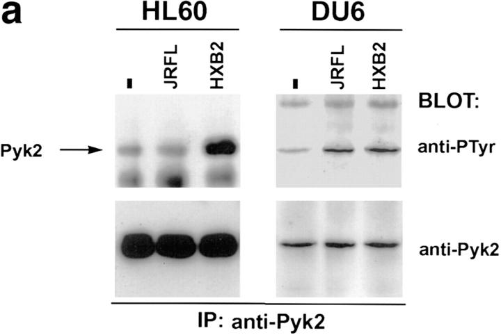
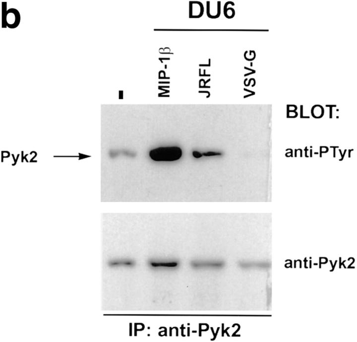
Figure 2.
Tyrosine phosphorylation of Pyk2 after contact with HIV-1 envelope glycoprotein on the surface of transfected 293T cells and virions. (a) HL60 or DU6 cells were lysed 30 s after being mixed with 293T cells expressing M-tropic (JRFL), T-tropic (HXB2), or no (−) envelope. (b) DU6 cells were mixed with HIV–luc particles pseudotyped with either JRFL or VSV-G envelopes and lysed after 90 s. As a positive control, MIP-1β was incubated with DU6 cells for 30 s before lysis.


