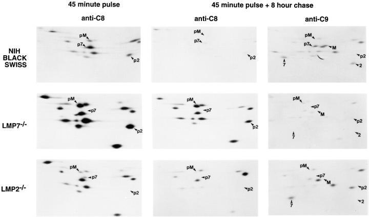
Figure 5.
Pulse-chase labeling of proteasomes synthesized in spleen cells from NIH Black Swiss (wild type), LMP7−/−, and LMP2−/− mice. Equal numbers of mouse spleen Con A–stimulated T cell blasts were labeled with [35S]methionine/cysteine for 45 min and then harvested (45 minute pulse) or chased for 8 h (45 minute pulse + 8 hour chase). At the end of each incubation, cells were lysed and proteasomes were immunoprecipitated from half of each lysate with anti-C8, which recognizes only 12-16S preproteasomes (anti-C8), and from the other half of each lysate with anti-C9, which recognizes 20S proteasomes and a small subset of 12-16S preproteasomes (anti-C9) (22). (Anti-C9 immunoprecipitates after a 45-min pulse demonstrated little radioactivity in 20S proteasomes and are not shown.) Proteasome subunits were separated by two dimensional NEPHGE-PAGE and visualized by autoradiography. Spots corresponding to inducible subunits and their precursors, as identified previously (22), are labeled (p2, pre-LMP2; 2, LMP2; p7, pre-LMP7; 7, LMP7; pM, pre-MECL; M, MECL). Note that pre-LMP7 is partially obscured by a larger spot (iota) in NIH Black Swiss preproteasomes. LMP7, LMP2, and their precursors have slightly different isoelectric points in NIH Black Swiss mice due to known polymorphisms (34). Immunoprecipitates from 5 × 10⁶ cells were loaded on each gel.

