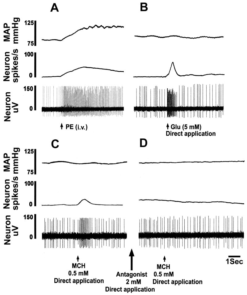
Fig. 5.
Excitation of mNTS neurons by MCH. In each panel, top trace: MAP (mmHg), 2nd trace: neuronal firing rate (spikes/sec), and 3rd trace: neuronal action potentials (μV). A: Basal firing rate of this neuron was 8 spikes/sec. Increase in MAP induced by phenylephrine (PE; 3 μg/kg, i.v.) elicited an increase in the neuronal activity (46 spikes/sec) indicating that the neuron was barosensitive. B: When the neuronal firing returned close to basal level (8 spikes/sec), pressure micro-application (4 nl) of L-Glu (5 mM) directly to the neuron increased its firing (58 spikes/sec) which lasted for about 0.8 sec. C: When the neuronal firing returned close to the basal level (10 spikes/sec), pressure micro-application (4 nl) of MCH (0.5 mM) increased the neuronal firing (28 spikes/sec) which lasted for about 0.8 sec. When the neuronal firing returned to the basal level (10 spikes/sec), MCH-1 receptor antagonist (2 mM) was applied (4 nl) (large arrow between panels C and D); no change in neuronal firing was elicited (not shown). D: Subsequent pressure micro-application (4 nl) of MCH (0.5 mM) to the neuron failed to elicit an increase in neuronal firing.

