Figure 1.
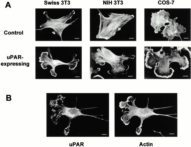
Effect of uPAR expression on cell morphology. (A) Cells in growth medium (DME plus 10% FCS or donor calf serum) were injected with the expression plasmid pRc/CMV-uPAR (100 μg/ml) or with FITC-dextran for controls. As an additional control, cells were injected with an empty expression vector, and these cells were identical to FITC-dextran–injected cells (data not shown). 4 h after injection, the cells were fixed, uPAR-expressing cells were identified by double immunofluorescence with a rabbit polyclonal anti–human uPAR (Nykjær et al. 1998) followed by FITC goat anti–mouse IgG, and the actin cytoskeleton was visualized with rhodamine-phalloidin. Typical morphologies of the actin cytoskeleton in uPAR-expressing and control cells in three different cell lines are shown. (B) Localization of ectopically expressed uPAR. Swiss 3T3 cells injected with pRc/CMV-uPAR were fixed and stained as described in A. The localization of uPAR and the organization of the actin cytoskeleton are shown. Bars, 10 μm.

