
Figure 5.
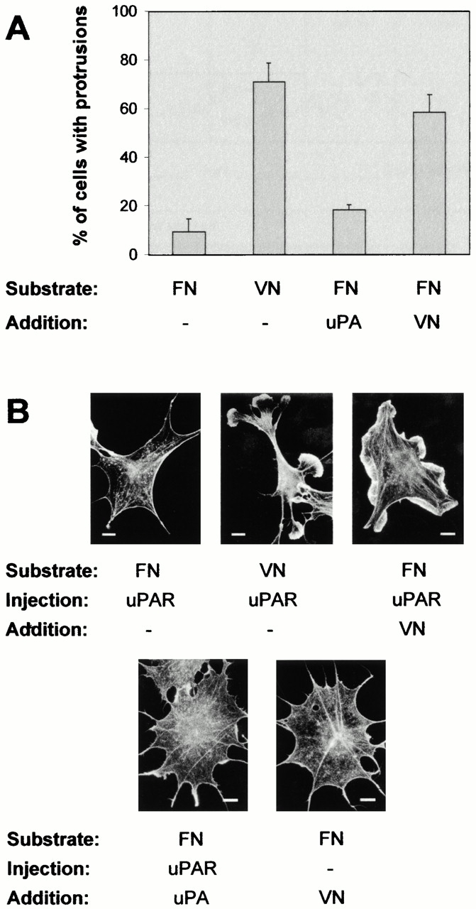
Role of extracellular matrix factors in uPAR-induced protrusions. (A) Growing Swiss 3T3 cells were trypsinized, washed two times in serum-free medium, and replated in serum-free medium on coverslips precoated with FN (50 μg/ml) or VN (10 μg/ml). 2 h after plating, cells were injected with pRc/CMV-uPAR. After 4 h of expression, cells were fixed, stained, and the number of uPAR-expressing cells with clearly identifiable protrusions/lamellipodia was determined as described above. In some cases, cells were treated with soluble VN (10 μg/ml) or DFP-uPA (10 nM) for 5 min in the presence of 0.05 mM cRGD before fixation. Data are average ± SD for at least three experiments, each examining 100 injected cells. (B) Cells plated on FN or VN as indicated were injected with pRC/CMV-uPAR, treated with soluble VN or DFP-uPA in the presence of 0.05 mM cRGD as indicated, then fixed and stained as described above. Typical morphologies of the actin cytoskeleton are shown. Bars, 10 μm.