
Figure 6.
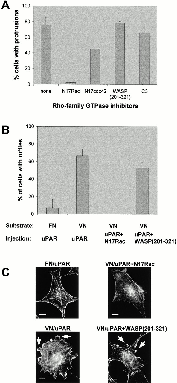
Role of Rho GTPases in uPAR-induced protrusions. (A) Swiss 3T3 cells in growth medium were coinjected with pRC/CMV-uPAR (100 μg/ml) and the expression plasmids pRK5–myc-N17Rac (20 μg/ml), pRK5–myc-N17cdc42 (100 μg/ml), or pRK5–myc-WASP(201–321) (40 μg/ml), or with 30 μg/ml of recombinant C3 transferase protein as indicated. After 4 h of expression, cells were fixed. uPAR expression was determined as described above. Expression of inhibitor constructs was verified by immunofluorescence staining with biotin-conjugated mouse anti-myc (clone 9E10) followed by Alexa-conjugated streptavidin. Data are average ± SD for at least three experiments, each examining 100 injected cells. (B) Subconfluent quiescent Swiss 3T3 cells plated on FN or VN were injected with pRC/CMV-uPAR (100 μg/ml) or coinjected with pRc/CMV-uPAR (100 μg/ml) and pRK5–myc-N17Rac (25 μg/ml) or pRK5–myc-WASP(201–321) (40 μg/ml) as indicated. After 3–8 h of expression, cells were fixed and stained as described above. The number of uPAR- or coexpressing cells with clearly identifiable lamellipodia was determined. Data are average ± SD for at least three experiments, each examining 100 injected cells. (C) Typical morphologies of quiescent, subconfluent Swiss 3T3 cells injected with expression plasmids as described in B. The morphology of the actin cytoskeleton as visualized by staining with rhodamine-phalloidin is shown. Typical lamellipodia are indicated by arrowheads. Bars, 10 μm.