
Figure 8.
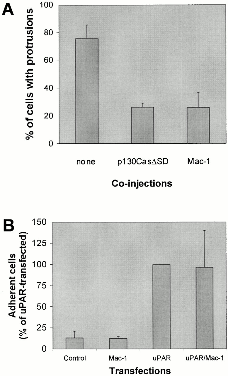
Effect of p130CasΔSD and Mac-1 on uPAR-induced protrusions. (A) Swiss 3T3 cells in growth medium were coinjected with pRC/CMV-uPAR (100 μg/ml) and either one of the expression plasmids pSSRα-p130CasΔSD/pEBG-GST-p130CasΔSD or both of the expression plasmids pRK5-CD11b and pRK5-CD18 (α- and β chain of Mac-1). After 4 h of expression, cells were fixed, stained, and the number of uPAR-expressing cells with clearly identifiable protrusions was determined as described above. Expression of inhibitor constructs was verified by immunofluorescence staining using anti-GST (rabbit polyclonal) or anti-CD11b (clone ICRF 44) as primary antibodies. Expression of untagged and GST-tagged p130CasΔSD gave identical results and the data were averaged for presentation. Data are average ± SD for at least three experiments, each examining 100 injected cells. (B) NIH 3T3 cells transfected with pEGFP-C1 and empty vector or pRC/CMV-uPAR and/or pRK5-CD11b and pRK5-CD18 as indicated were subjected to detachment assays (see Materials and Methods for details) in the presence of 5 mM EDTA. Results are expressed as the fraction of adherent cells relative to the number uPAR-transfected adherent cells under these conditions. Results are mean ± SD of three experiments each performed in triplicate.