Figure 2.
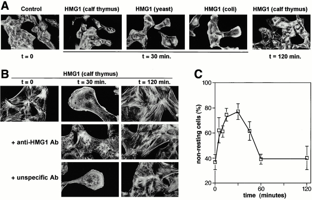
Effect of HMG1 on RSMC morphology and actin cytoskeleton organization. (A) Subconfluent (50–70%) cultures of RSMC were challenged for the indicated times with HMG1 (100 ng/ml), either purified from calf thymus or expressed in yeast or E. coli as indicated. Actin filaments were visualized using TRITC-phalloidin. (B) Anti–HMG1 rabbit antibodies, but not unspecific rabbit antibodies, inhibit HMG1-stimulated cytoskeleton reorganization. RSMC were pretreated overnight with either anti–HMG1 (2 μg/ml) or unspecific control antibodies (2 μg/ml), and then 100 ng/ml HMG1 (from calf thymus) was added. (C) RSMC were stimulated with 100 ng/ml HMG1. Quantification of the actin cytoskeleton reorganization was performed by taking low-magnification photographs and counting the cells in each state of cytoskeleton organization. Resting cells (state 1) exhibit numerous stress fibers. Nonresting cells (state 2) show a reorganization of actin cytoskeleton: a decrease of stress fibers content, membrane ruffling, actin semi-ring with an elongated polarized morphology characteristic of motile RSMC.

