
Figure 2.
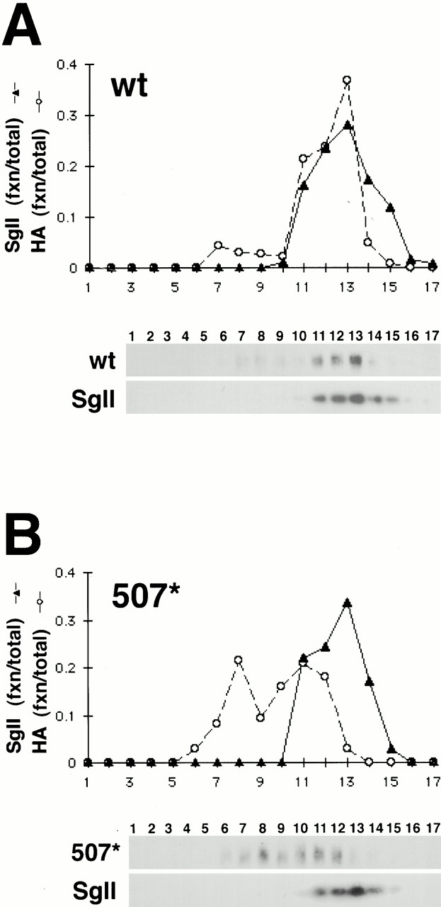
Density gradient fractionation of wild-type and 507* VMAT2. Post-nuclear supernatants from PC12 cells stably expressing HA-tagged wild-type (A) and 507* (B) VMAT2 were separated by equilibrium gradient centrifugation through 0.6–1.6 M sucrose. Fractions were collected and analyzed by Western blot using the monoclonal HA antibody to detect VMAT2 (A and B, top), and a polyclonal antibody to detect the LDCV marker SgII (bottom). The immunoblots were then digitized and quantified using NIH Image. For each fraction, the amount of VMAT2 and SgII is expressed as a percentage of total immunoreactivity in all the fractions. Wild-type VMAT2 cofractionates with SgII in heavy fractions, whereas 507* cofractionates to a lesser extent with SgII and appears instead in lighter fractions. The analysis of three different stable cell lines for each construct yielded similar results.