
Figure 3.
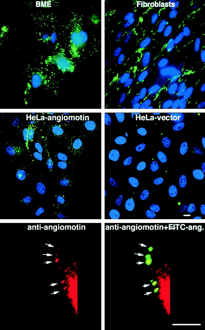
Binding of FITC-labeled angiostatin to living cells in culture. Cells were incubated with 10 μg/ml FITC-labeled angiostatin at 4°C for 60 min. The cells were placed in 37°C incubator 15 min before washing in PBS and fixation in 3.7% formaldehyde. Angiostatin binds to and becomes internalized in bovine microcapillary endothelial (BME) cells resulting in a patchy staining of endosomes (top left). In contrast, angiostatin binds to the matrix of human fetal fibroblasts but is not internalized (top right). HeLa cells transfected with angiomotin bind and internalize angiostatin in a similar pattern to that of endothelial cells (middle left). HeLa-Ctrl cells are transfected with an empty vector and were negative for angiostatin binding (middle right). The bottom panel shows colocalization of FITC-angiostatin (ang.) with angiomotin during internalization after surface binding to HeLa cells transfected with angiomotin. Angiomotin was visualized by immunofluorescent staining using immunoaffinity-purified rabbit polyclonal antibodies against angiomotin as described in Materials and Methods. Bars, 10 μm.