
Figure 5.
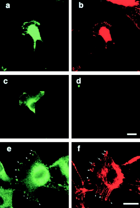
Subcellular localization of transfected and endogenous angiomotin. GFP-tagged angiomotin was expressed in NIH 3T3 cells using a retroviral expression system. Colocalization of GFP-angiomotin (a) with FAK (b) in the extending lamellipodium. (c) Immunofluorescence staining with a polyclonal antibody against angiomotin shows similar localization of endogenous angiomotin in a migrating HUVEC. (d) Control IgG from the same immunized animal. e and f shows the localization of angiomotin (e) to actin ruffles (f) and focal complexes in spreading HUVECs as visualized by phalloidin staining (arrows). Bars, 10 μm.