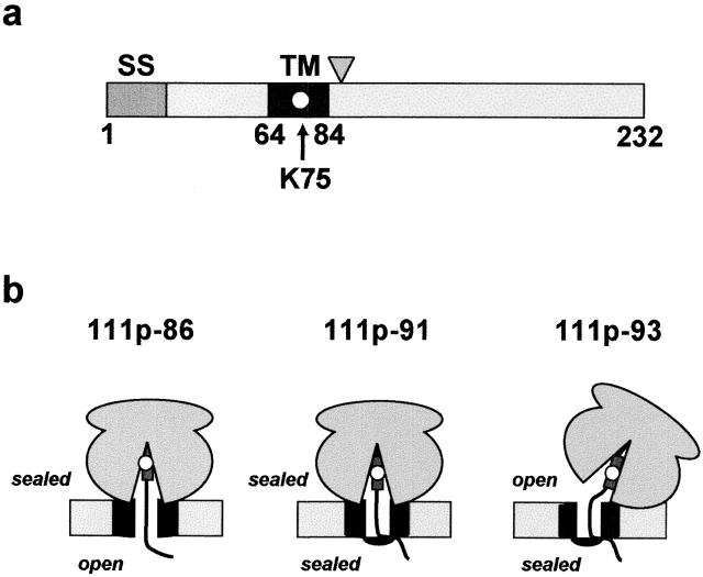
Figure 2.
111p protein and integration intermediates. (a) The chimeric 111p single-spanning membrane protein was constructed using the TM sequence from vesicular stomatitus G protein (residues 65–84, dark gray) and lysine-free stretches of preprolactin and Bcl-2 (Liao et al., 1997). 111p contains only a single lysine codon at position 75 in the middle of the TM sequence (white circle). Note that the TM segment will still be nonpolar and uncharged when ɛNBD-Lys is incorporated at this location. The signal sequence (SS) is indicated in gray (residues 1–22), and the triangle indicates the approximate position of the truncations used in this study (residues 86, 91, and 93). (b) Accessibility of the fluorescent probe from either the cytoplasmic or lumenal sides of intact ER microsomes is indicated for each intermediate used in this study (Liao et al., 1997). The ribosome is shown bound to the translocon (black) at the ER membrane (light gray) in each case. The TM sequence is represented by a dark gray rectangle and the fluorescent probe by a white circle.

