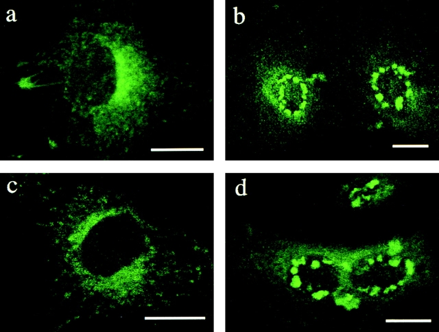
Figure 4.
Increased expression of PLP changes the normal distribution of BODIPY–lactosylceramide and –galactosylceramide. (a–d) BHK cells were left uninfected (a and c) or were infected with SFV-PLP–myc for 16 h (b and d). BODIPY-labeled lipids were added to the cells as BSA complexes for 1 h at 37°C, and then lipids were washed away and internalization was allowed to proceed for 90 min. After “back exchange” (see Materials and methods) of the lipids at 12°C, the distribution of the lipid was monitored. Distribution of BODIPY–lactosylceramide is shown in uninfected cells (a) and infected cells (b). Distribution of BODIPY–galactosylceramide is shown in uninfected (c) and infected cells (d). Bars, 10 μm.

