Figure 2.
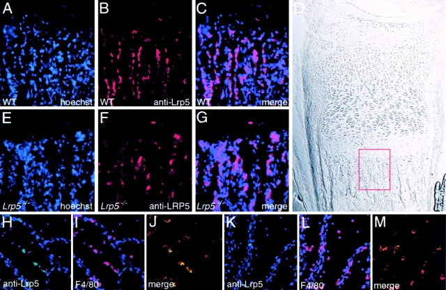
Lrp5 expression in osteoblasts and ocular macrophages. (A–C) Lrp5 expression in osteoblasts and bone lining cells in the primary spongiosa of the femur in wild-type mice. (A) Hoechst nuclear staining is shown in blue; (B) Anti-Lrp5 immunoreactivity in red. (C) A merge of the red and blue channels. (D) DIC image of distal femur from wild-type with a box showing the region examined in A–C. (E–G) Lrp5 expression in the primary spongiosa of the femur in Lrp5 − / − mice. (E) Hoechst nuclear staining. (F) Anti-Lrp5 immunoreactivity. (G) Merge. (H–M) Whole-mount preparations of hyaloid vessels from wild-type (H–J) and Lrp5 − / − (K–M) mice. (H and K) Immunoreactivity for Lrp5. (I and L) Immunoreactivity with the macrophage-specific antibody F4/80, both with Hoechst nuclear staining. (J and M) Merge of the red, blue, and green channels; yellow cells indicate that macrophages are positive for Lrp5.

