
Figure 4.
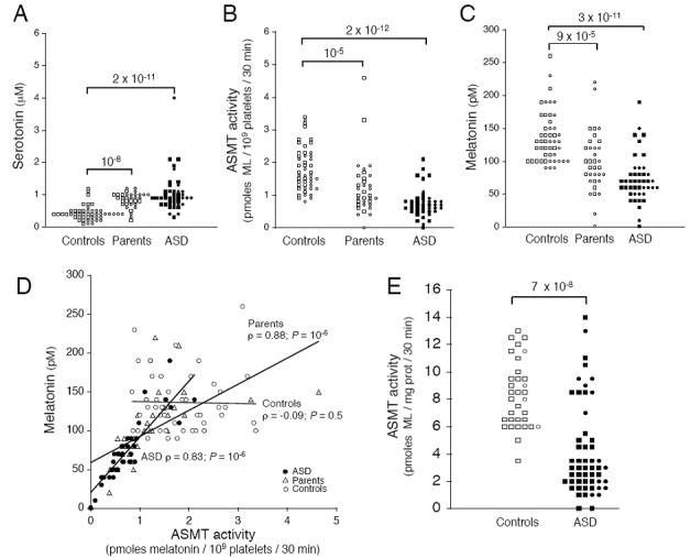
ASMT activity and melatonin concentration in patients, parents and controls. (a) Serotonin levels, measured in the platelets of 43 ASD patients (mean±s.d.; 1±0.6 μM), 34 parents (0.8±0.2 μM) and 48 controls (0.4±0.2 μM). (b) ASMT activity, measured in the platelets of 43 ASD patients (0.73±0.43 pmol/109 platelets/30 min), 34 parents (1.20±0.85 pmol/109 platelets/30 min) and 48 controls (1.81±0.68 pmol/109 platelets/30 min). (c) Plasma melatonin concentration in 43 autism spectrum disorders (ASD) patients (73±36 pmol), 34 parents (99±46 pmol) and 48 controls (136±39 pmol). (d) Blood melatonin concentration expressed with respect to ASMT activity in platelets. ASMT activity and melatonin concentration were not correlated in controls (white circles). In contrast, in ASD patients (black circles), and their parents (white triangles), a significant correlation was observed. Spearman’s rho test was used to evaluate the correlation between ASMT activity and melatonin concentration, (e) ASMT activity in B lymphoblastoid cell lines from 53 ASD patients (3.9±3.4) and 33 controls (8.3±2.4). Circles and squares indicate female and male subjects, respectively. Statistical significance was assessed with the Mann–Whitney U-test.