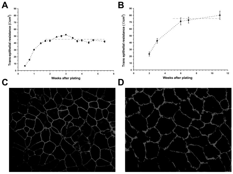
Figure 1.
Resistance and morphology of RPE cell monolayers. A, Increase in transepithelial resistance of ARPE-19 cell monolayers following cell plating on transwell inserts. The dashed line represents the average resistance (45 ± 4 Ωcm2) for confluent monolayers between weeks 2 and 5. Cultures were tested for up to 8 weeks; however, routine experiments were performed following three weeks in culture. B, Increase in transepithelial resistance of porcine primary RPE cell monolayers following cell plating on transwell inserts. The dashed line represents the average resistance (76 ± 9 Ωcm2) achieved for confluent monolayers between weeks 5 and 11. Values are means ±SE. Immunohistochemistry of confluent ARPE-19 cells (C) and porcine primary RPE cells (D) with mouse anti-ZO-1 as primary, and FITC-conjugated anti-mouse as secondary antibodies. The magnification was ×200.

