
Figure 1.
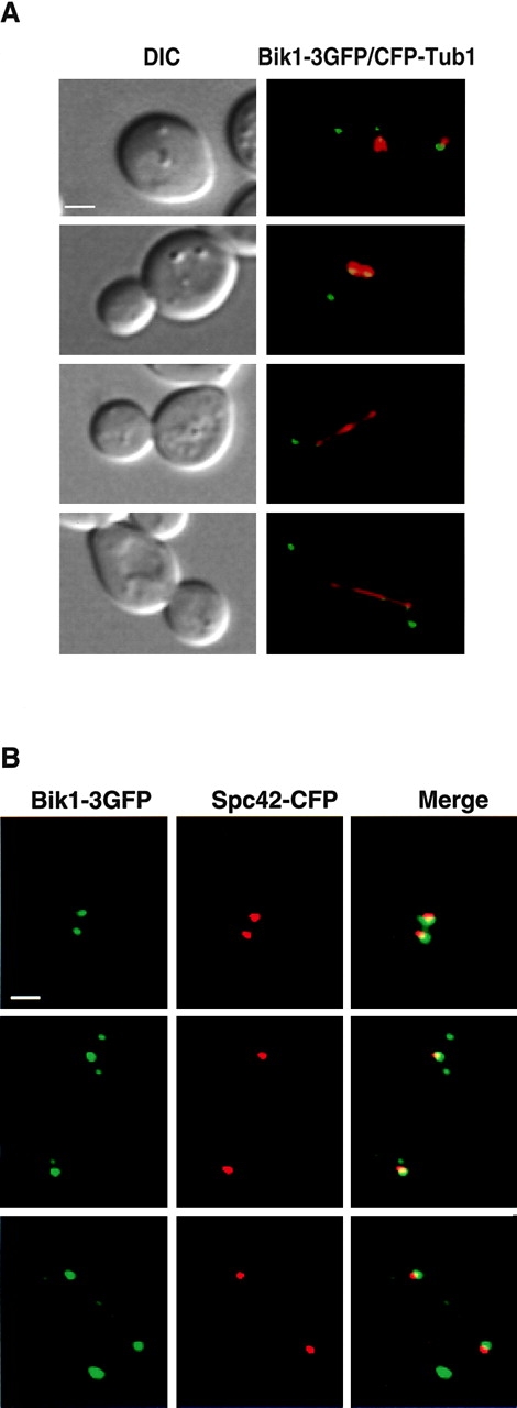
Localization of Bik1 to MT plus ends and to the kinetochore. (A) Colocalization of Bik1–3GFP (green) with CFP-Tub1 (red) at different stages of the cell cycle. Pairs of DIC (left) and merged fluorescence (right) images are shown. From top to bottom: G1 cell; preanaphase cell; anaphase cell; and telophase cell. Bar, 2 μm. (B) Bik1–3GFP localization at the kinetochore during mitosis in cells expressing Bik1–3GFP (green) and Spc42-CFP (red), a spindle pole body marker. Bar, 2 μm.